Coeliac disease: investigation
Coeliac disease is caused by sensitivity to the protein gluten. Repeated exposure leads to villous atrophy which in turn causes malabsorption. Conditions associated with coeliac disease include dermatitis herpetiformis (a vesicular, pruritic skin eruption) and autoimmune disorders (type 1 diabetes mellitus and autoimmune hepatitis).
Diagnosis is made by a combination of immunology and jejunal biopsy. Villous atrophy and immunology normally reverses on a gluten-free diet.
NICE issued guidelines on the investigation of coeliac disease in 2009. If patients are already taking a gluten-free diet they should be asked, if possible, to reintroduce gluten for at least 6 weeks prior to testing.
Immunology
Jejunal biopsy
Rectal gluten challenge has been described but is not widely used
Diagnosis is made by a combination of immunology and jejunal biopsy. Villous atrophy and immunology normally reverses on a gluten-free diet.
NICE issued guidelines on the investigation of coeliac disease in 2009. If patients are already taking a gluten-free diet they should be asked, if possible, to reintroduce gluten for at least 6 weeks prior to testing.
Immunology
- tissue transglutaminase (TTG) antibodies (IgA) are first-choice according to NICE
- endomyseal antibody (IgA)
- anti-gliadin antibody (IgA or IgG) tests are not recommended by NICE
- anti-casein antibodies are also found in some patients
Jejunal biopsy
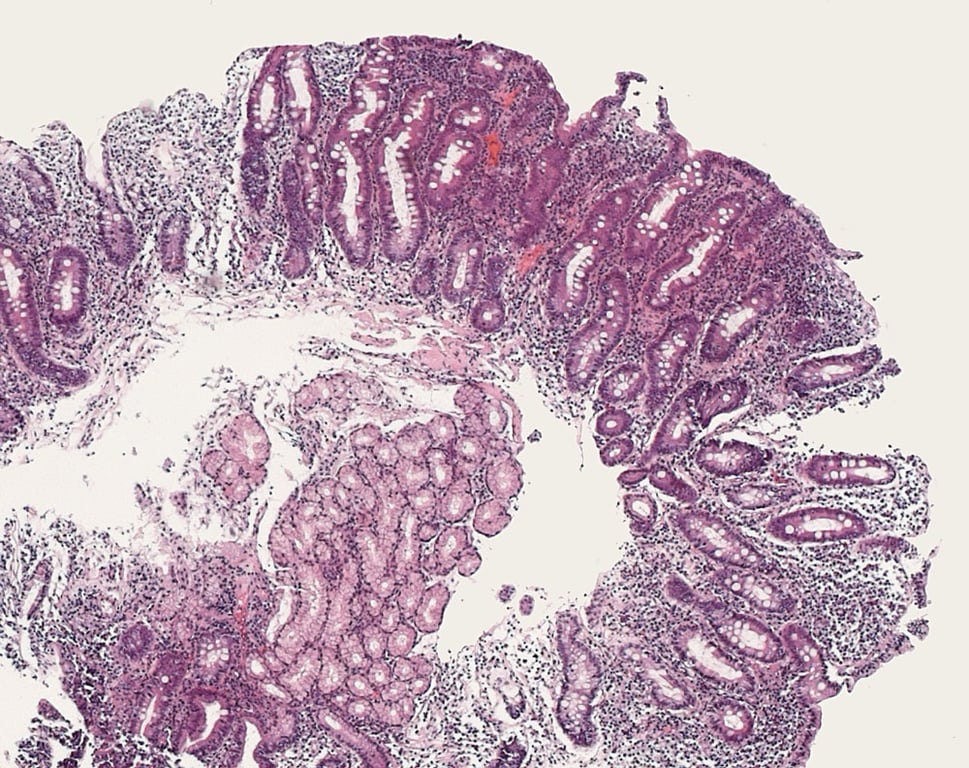
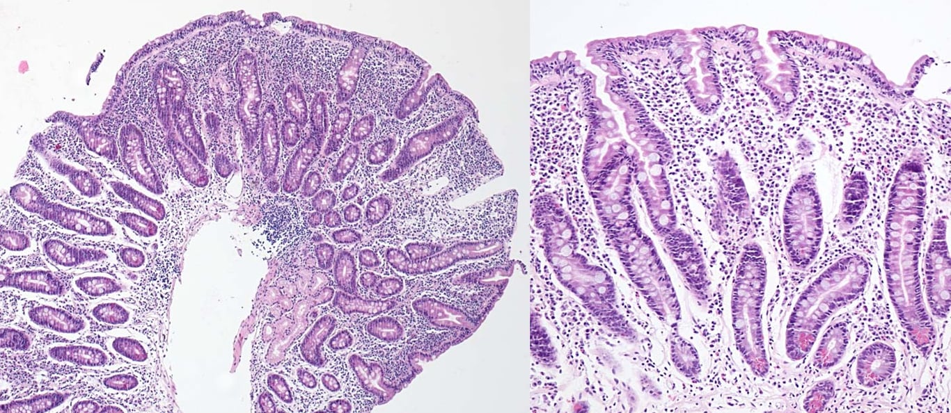
- villous atrophy
- crypt hyperplasia
- increase in intraepithelial lymphocytes
- lamina propria infiltration with lymphocytes
Rectal gluten challenge has been described but is not widely used
 | |
| © Image used on license from PathoPic | |
Duodenal biopsy from a patient with coeliac disease. Complete atrophy of the villi with flat mucosa and marked crypt hyperplasia. Intraepithelial lymphocytosis. Dense mixed inflammatory infiltrate in the lamina propria.
 | |
| © Image used on license from PathoPic | |
Duodenal biopsy from a patient with coeliac disease. Flat mucosa with hyperplastic crypts and dense cellular infiltrate in the lamina propria. Increased number of intraepithelial lymphocytes and vacuolated superficial epithelial cell vacuolated superficial epithelial cells. Higher magnification image on the right.