Cardiovascular physiology: filtration, lymphatics and oedema
On this page
Filtration
The capillary wall is a semi-permeable membrane dividing the intravascular and interstitial compartments that acts as a filter.Permeability varies for different types of molecules. Permeability is much greater for smaller molecules such as water and electrolytes than it is for larger molecules such as proteins.
The exchange of fluids across the capillary membrane is primarily governed by oncotic (colloid osmotic) and hydrostatic pressures. There are components of each on either side of the capillary wall leaving four pressure components as shown below:
- Capillary hydrostatic pressure
- Tissue (interstitial) hydrostatic pressure
- Capillary (plasma) oncotic pressure
- Tissue (interstitial) oncotic pressure.
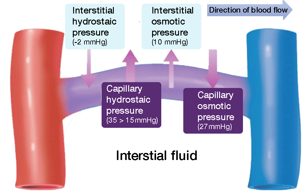 | |
Diagram illustrating pressure components across the capillary. Note how capillary hydrostatic pressure varies from around 35mmHg at the arteriolar end to 15mmHg at the venule end
There is an overall net flow depending on the pressures. The oncotic pressures are consistent along the course of the capillary. Within the capillary oncotic pressure is typically around 27 mmHg whilst in the interstitial fluid it is between 5-10mmHg. This leaves an overall oncotic pressure difference of 17-22 mmHg which acts to drive water into the capillary.
For hydrostatic pressure the direction of net flow changes during the course of the capillary due to the change in capillary hydrostatic pressure from the arterial end to the venous end of the capillary. Interstitial hydrostatic pressure can be considered negligible at close to zero. Capillary hydrostatic pressure is 35mmHg at the arterial end of the capillary which is greater than the 17mmHg net oncotic pressure acting against it so the overall effect is movement of water out of the capillary (filtration) at this end of the capillary. At the venous end of the capillary capillary hydrostatic pressure has dropped to around 15mmHg which is less than the 17-22mmHg oncotic pressure so flow reverses allowing for resorption to occur. Overall there is net filtration of around 2 litres of fluid per day.
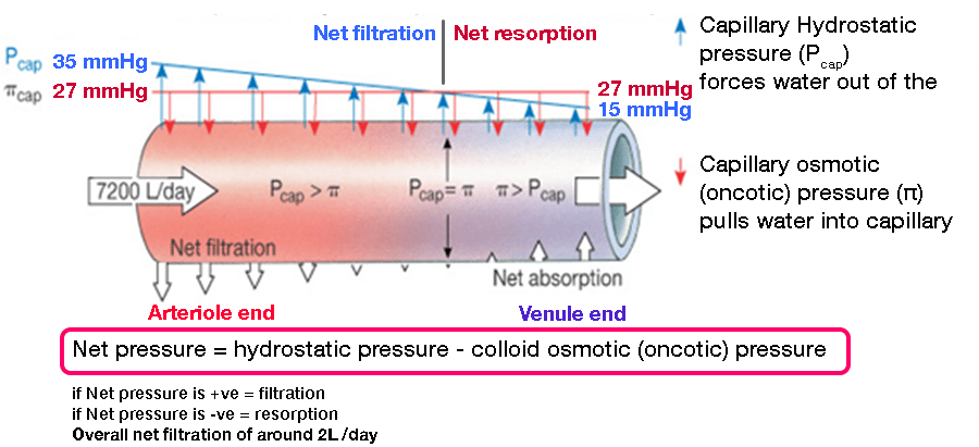 | |
Interstital hydrostatic and interstitial oncotic pressures are sometimes considered negligible as it is the capillary oncotic and capillary hydrostatic forces that are the main determinants of flow across the capillary membrane. Note how the oncotic pressure is constant whereas the hydrostatic pressure drops across the length of the capillary. Click on image to enlarge
Remember:
Water flows from low to high oncotic pressureWater flows from high to low hydrostatic pressure
It can be helpful to think of oncotic pressure as a pulling force (towards proteins) and hydrostatic pressure as a pushing force.
 | |
| Wikipedia | |
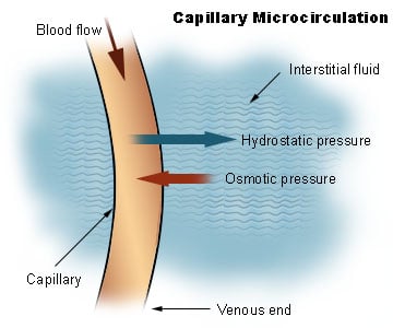
Diagram illustrating net movements in and out of capillary Click on image to enlarge
Oncotic pressure
Oncotic pressure (AKA colloid osmotic pressure) is pressure induced by proteins, most notably albumin in a blood vessel's plasma. Proteins displace water and create a relative water deficit which draws water towards them
Hydrostatic pressure
Hydrostatic pressure in blood vessels is the pressure of the blood against the wall. It is the opposing force to oncotic pressure.The overall net movement of water is determined by the balance between the hydrostatic and oncotic pressures.
It may be represented by Starling's equation below:
Where:
It may be represented by Starling's equation below:
- net flow of water ˆ (Pc - Pi) - σ(Ï€ p-Ï€ i)
Where:
- Pc - Pi is the net hydrostatic pressure
- π p-π i is the net oncotic pressure
- σ is the reflection coefficient (a measure of protein permeability)
Lymphatics
Fluid filtered by the microcirculation returns to the blood circulation via the lymphatics.
In an average adult approximately 8L of fluid per day are filtered by the microcirculation and returned to the lymphatics
Lymphatic capillaries are blind ending bulbous tubes that permit the entry of fluid, proteins and bacteria but do not allow their exit.
Lymphatic capillaries merge to form collecting lymphatics and larger afferent lymph vessels. Movement of lymph is facilitated by valves to prevent the backflow of lymph (similar to veins) and smooth muscle which contracts to push lymph through the lymphatic system. The lymph is driven to nodes where phagocytes remove foreign material such as bacteria.
At the lymph nodes some fluid is resorbed through capillaries whilst the rest leaves the node via efferent lymph vessels and is returned to the circulation via the thoracic duct.
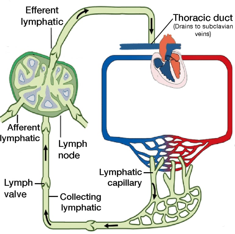 | |
Overview of lymphatic system
Oedema
Oedema is swelling of the tissues as a result of excessive fluid within the interstitial space.
Oedema occurs when filtration exceeds interstitial fluid removal by the lymphatics
Summary of oedema causes
| Mechanism | Causes |
|---|---|
| Increased filtration | Increased venous pressure:
|
| Obstruction to lymphatic drainage |
|
| Change in capillary permeability |
|