Cerebral ventricles
The ventricular system of the brain consists of four interconnected cavities called cerebral ventricles. The ventricular system is continuous with the central canal of the spinal cord communicating with the spinal cord via the fourth ventricle.
Each ventricle contains specialised cells called the choroid plexus which produces cerebrospinal fluid (CSF). The ventricular system and the central canal of the spinal cord are lined with another type of specialised epithelial cell, called ependyma, that are connected by tight junctions which maintain the blood-cerebrospinal fluid barrier.
The ventricles are connected by channels called foramina. CSF flows between the ventricles via these foramina. The interventricular foramina (AKA foramina of Monro) connects the lateral ventricles to the third ventricle whilst the cerebral aqueduct (aqueduct of Sylvius) connects the third and fourth ventricles. the foramina are summarised in the table below:
Each ventricle contains specialised cells called the choroid plexus which produces cerebrospinal fluid (CSF). The ventricular system and the central canal of the spinal cord are lined with another type of specialised epithelial cell, called ependyma, that are connected by tight junctions which maintain the blood-cerebrospinal fluid barrier.
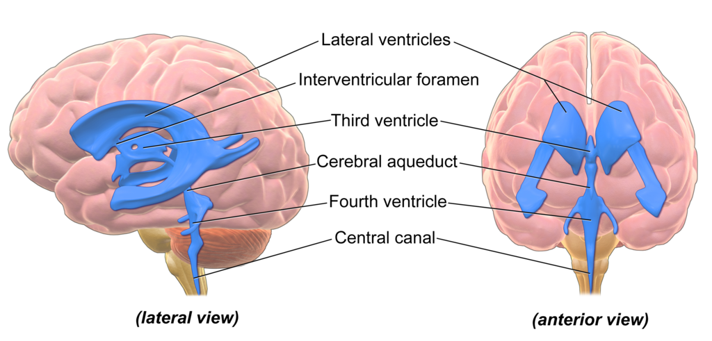 | |
Illustration of the anatomy of the cerebral ventricles. Click on image to enlarge
The ventricles are connected by channels called foramina. CSF flows between the ventricles via these foramina. The interventricular foramina (AKA foramina of Monro) connects the lateral ventricles to the third ventricle whilst the cerebral aqueduct (aqueduct of Sylvius) connects the third and fourth ventricles. the foramina are summarised in the table below:
| Foramina | Origin | Destination |
|---|---|---|
| Interventricular foramina (Monro) | Lateral ventricles | Third ventricle |
| Cerebral aqueduct (Sylvius) | Third ventricle | Fourth ventricle |
| Median aperture (Magendie) | Fourth ventricle | Subarachnoid space via cisterna magna |
| Right & left lateral aperture (Luschka) | Fourth ventricle | Subarachnoid space via cistern of great cerebral vein |