Cardiovascular physiology: Cardiac conduction & the cardiac cycle
On this page
Conduction pathway of the heart
 | |
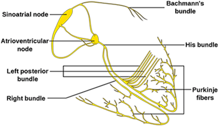 | |
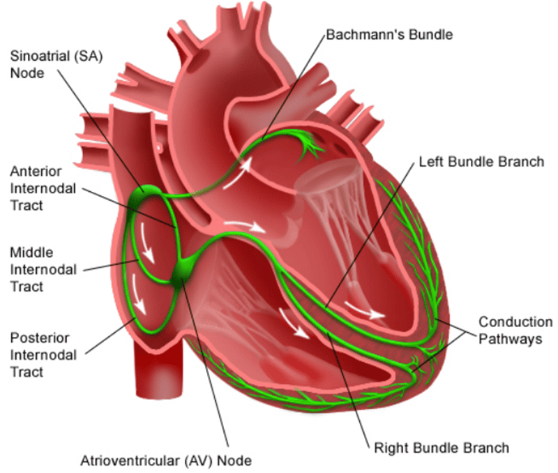
Above show schematics of the conduction system of the heart. Normally depolarisation is initiated at the SA node and spreads via nerve fibres through both atria before reaching the AV node and then entering the bundle of His before passing down the left and right bundle branches and Purkinje fibres
Sinatrial node
The SA node initiates the cardiac cycle. Impulses cause a depolarization wave that spreads through the atria. Gap junctions permit the spread of depolarization. The annulus fibrosus prevents direct spread of depolarization to the ventricles. As a result depolarization is chanelled via the AV node.Atrioventricular node
The AV node is located in the posterior septal wall of the right atrium near the coronary sinus close to the atrial septum. It is composed of small cells and slows conduction of electrical impulses by around 120-200ms. This allows time for the atria to complete contraction and complete ventricular filling before the ventricles contract. On the ECG this is seen as the PR interval.Propagatory fibres
As the ventricles complete filling the electrical impulse from the AV node reaches the bundle of His which carries the depolarisation wave to the left and right bundle branches and the Purkinje fibres.Pacemaker & Pacemaker potential
In healthy individuals the heart rate is dictated by the SA node with rates between 60-100 bpm.The SA node lacks fast Na channels
The SA node lacks fast Na channels. Depolarisation of the SA node myocytes is via influx of Ca2+ ions via slow Ca2+ channels (rather than influx of sodium via fast Na channels). This results in slower depolarisation.
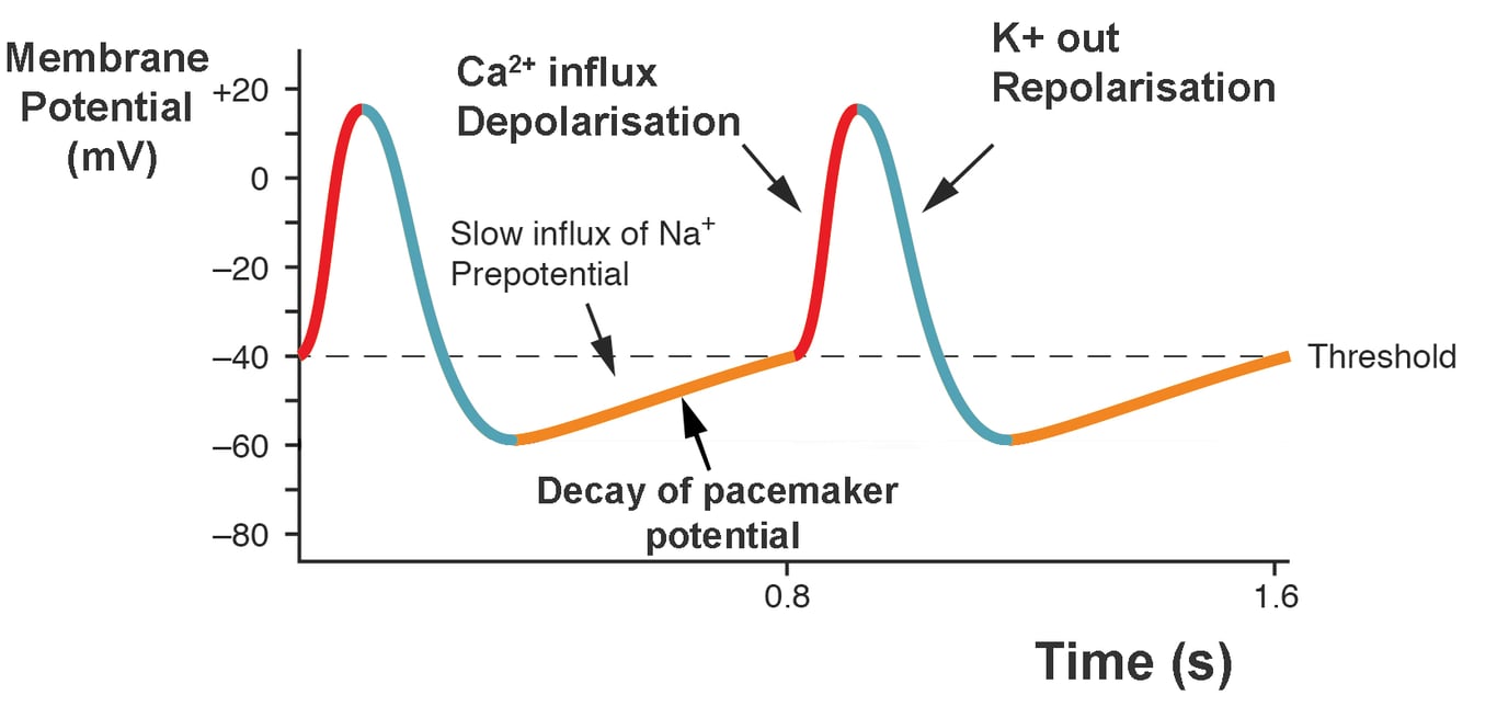 | |
The pacemaker potential is the slow increase in voltage across the cell's membrane (the membrane potential) that occurs between the end of one action potential and the beginning of the next action potential (highlighted orange on the image above). This increase in membrane potential is what causes the cell membrane, which typically maintains a resting membrane potential of -60 to -70 mV to reach the threshold potential and consequently fire the next action potential. The pacemaker potential is what drives the self-generated rhythmic firing (automaticity) of pacemaker cells. The rate of decay of the pacemaker potential is what determines the timing of the next action potential and the heart rate.
If the SAN fails e.g. conduction block then the AVN will typically take over pacing but its rate is typically 40-60 bpm. If both the SAN and AVN fail pacing will be initiated in the ventricles with rates around 20-40 bpm.
The ECG and the cardiac cycle
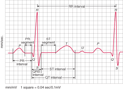 | |
Image illustrating ecg intervals
Key points
It should be noted that atrial repolarisation does occur at the time of the QRS however the QRS complex itself is due to ventricular depolarisation.
- P wave = Atrial depolarisation
- QRS complex = Ventricular depolarisation
- T wave = Ventricular repolarisation
- U wave = Repolarisation of the interventricular septum
It should be noted that atrial repolarisation does occur at the time of the QRS however the QRS complex itself is due to ventricular depolarisation.