Thoracic inlet
The thoracic inlet is an important space due to the structures within it. Knowledge of the relations of oesophagus, trachea, aortic arch, subclavian arteries and veins and brachiocephalic trunk within the thoracic inlet are required knowledge.
The diagram below shows the arrangements of these structures at the thoracic inlet (also known as the superior thoracic aperture)
Diagram illustrating the structures passing through the thoracic Inlet
The key structures passing through the inlet are:
Viscera:
Boundaries
- Located at T1 level and is bounded by T1 posteriorly
- Lateral boundaries are 1st ribs and costal cartilages
- Anterior boundary is the superior border of the manubrium
The diagram below shows the arrangements of these structures at the thoracic inlet (also known as the superior thoracic aperture)
Diagram illustrating the structures passing through the thoracic Inlet
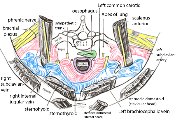 | |
Note the position of the first ribs as these are the lateral borders. Not all of the structures labelled pass through the inlet, some such as the brachial plexus are closely related
The key structures passing through the inlet are:
Viscera:
- Trachea
- Oesophagus
- Lung apicies
- Thymus
- Phrenic nerve
- Vagus nerve
- Recurrent laryngeal nerves
- Sympathetic trunks
- Common carotid arteries
- Subclavian arteries
- Internal jugular veins
- Brachiocephalic veins
- Subclavian veins
- Thoracic duct
- Lymph nodes and lymphatic vessels