Immunoglobulins (antibodies)
On this page
Production and distribution
Immunoglobulins are produced by plasma cells (differentiated B lymphocytes) in the following tissues:- Bone marrow
- Lymph nodes
- Spleen
- Mucosa-associated lymphoid tissue (MALT) e.g. Peyer’s patches, tonsils
Once produced antibodies may be found in:
- Serum (blood/plasma): mainly IgG, IgM
- Mucosal secretions: IgA (coats epithelial surfaces in GI, respiratory, genitourinary tracts)
- Bound to cells via Fc receptors (IgE on mast cells, basophils & eosinophils, IgG on macrophages, neutrophils & NK cells)
- On B-cell surfaces: IgM and IgD act as antigen receptors
Structure
Immunoglobulins (antibodies) are composed of 2 identical heavy chains and 2 identical light chains joined by disulphide (disulfide) bonds.
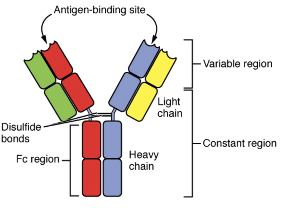 | |
Antibody structure. Click on image to enlarge
Heavy chains determine isotype: γ (IgG), α (IgA), μ (IgM), ε (IgE), δ (IgD).
Light chains are either κ (kappa) or λ (lambda).
Variable region (of heavy and light chains): gives antigen specificity.
Constant region: defines class and effector functions.
Fab region: antigen binding.
Fc region: binds complement and Fc receptors on immune cells.
Hinge region: provides flexibility.
Antibody forms:
- IgA: monomer (serum) or dimer (secretions).
- IgM: pentamer (serum) or monomer (B-cell receptor).
- IgD, IgE and IgG: monomers.
Types
| Class | Structure | Serum % | Main functions |
|---|---|---|---|
| IgG | Monomer | ~75–80% | Main antibody in secondary response; crosses placenta; opsonisation; complement activation; ADCC |
| IgA | Monomer (serum), dimer (secretions) | ~15% | Mucosal immunity (respiratory, GI, saliva, tears, breast milk); passive protection in infants |
| IgM | Pentamer (serum), monomer (BCR) | ~10% | First antibody in primary response; strong complement activator; B-cell receptor (monomeric form) |
| IgE | Monomer | <1% | Mast cell/basophil degranulation; allergic reactions; defence against helminths |
| IgD | Monomer | <1% | Present on naïve B-cells as receptor; role largely unknown |
Functions
- Neutralisation: toxins, viruses, prevent pathogen adherence.
- Opsonisation: tag antigens for phagocytosis.
- Agglutination: clumping antigens for clearance.
- Complement activation (classical pathway → lysis, opsonisation).
- ADCC: antibody-dependent cell-mediated cytotoxicity (via NK cells, cytotoxic T cells).
- B-cell receptor function (IgM, IgD).
- Mucosal immunity (IgA-mediated).
- Mast cell/basophil degranulation (IgE, also some IgG).
- Passive immunity to newborn (IgG via placenta, IgA via breast milk).