Erythrocyte Sedimentation Rate (ESR)
On this page
Background
Erythrocyte sedimentation rate (ESR) is an indirect, non-specific marker of inflammation. ESR measures the rate at which red blood cells sediment in a vertical tube over 1 hour. Normally, RBCs fall slowly, leaving little clear plasma.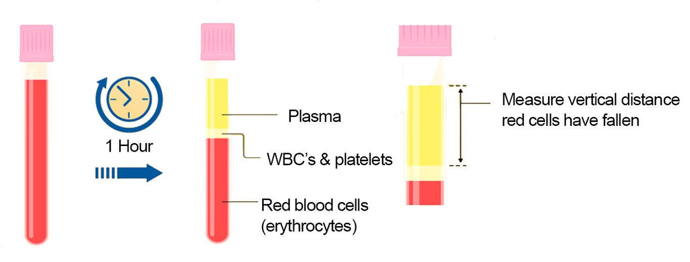 | |
Erythrocyte sedimentation rate (ESR. Click on image to enlarge
Principle (mechanism)
Under normal conditions red blood cells (RBCs or erythrocytes) carry a negative surface charge, primarily due to sialic acid residues on glycoproteins. The negative charge, referred to as zeta potential, causes red cells to repel each other.
In inflammation, acute-phase proteins (mainly fibrinogen, but also CRP and immunoglobulins) rise. These positively charged (or less negatively charged) proteins may bind to sialic acid residues and reduce the negative charge reducing the repulsive force between cells. Proteins such as fibrinogen can also bind multiple RBCs forming cross-linked bridges which also promotes RBC aggregation. These actions promote rouleaux formation (stacking).
Rouleaux formation refers to the way red blood cells (RBCs) stack together in linear chains, resembling a stack of coins, when suspended in plasma.
Rouleaux are denser and sediment more quickly (larger aggregates have a higher settling velocity as described by Stokes' law) leading to higher ESR.
Clinical uses
ESR is a non-specific marker: does not identify cause, but reflects inflammatory burden. Particularly useful in diagnosing and monitoring:- Temporal arteritis (giant cell arteritis)
- Polymyalgia rheumatica
- Some other autoimmune diseases and chronic infections.
Causes of raised ESR
- Inflammation and infection
- Anaemia (fewer RBCs, faster fall)
- Renal impairment
- Pregnancy
- Old age
- Monoclonal gammopathies e.g., multiple myeloma, Waldenström’s macroglobulinaemia (very high ESR even without inflammation)
Causes of low ESR
- Polycythemia (more RBCs, slower fall)
- Extreme leukocytosis (interference with sedimentation)
- Certain protein abnormalities
ESR vs CRP
- ESR: rises/falls slowly (days–weeks), reflects chronic inflammation.
- CRP: rises/falls rapidly (hours), better for acute monitoring.
Normal ranges
The normal range varies by NHS trust and laboratory. As ESR is higher in older adults many trusts have age dependent ranges whilst others do not. Some example reference ranges are shown below:
North West London NHS Pathology ESR normal range
- 0-17mm/hr for Males
- 0-23mm/hr for Females
University Hospital North Midlands ESR normal range
| Age | Female | Male |
|---|---|---|
| <16 years | 1-8 mm/hr | 1-8 mm/hr |
| 16-60 years | 2-16 mm/hr | 2-10 mm/hr |
| >60 years | 2-20 mm/hr | 2-18 mm/hr |
Key points
- ESR is sensitive but non-specific
- It is strongly influenced by plasma proteins, RBC count/shape, and patient factors such as age and pregnancy
- Very high ESR is classic in temporal arteritis, polymyalgia rheumatica, and paraproteinaemias