Calcium balance
On this page
99% of body calcium is found in the skeleton as hydroxyapatite. The remaining 1% is predominantly found in extracellular fluid.
In the plasma the distribution of calcium is approximately as follows:
Renal calcium handling
Calcium that is not protein bound, i.e. free ionised calcium and calcium salts, are freely filtered at the glomerulus. Overall over 95% of filtered calcium is reabsorbed along the nephron.
Filtered calcium is reabsorbed passively in the proximal tubule and loop of Henle along its electrochemical gradient created by sodium and water reabsorption.
Approximately 65-70% is reabsorbed in the proximal tubule and approximately 20-25% is reabsorbed in the thick ascending limb of the loop of Henle.
Up to 10% of the ultrafiltered calcium reaches the DCT and connecting tubule (DCT-CNT). The connecting tubules are a small segment between the distal tubule and cortical collecting tubule. Calcium transport is actively regulated according to calcium levels. Reabsorption of calcium is summarised in the box below. Typically around 8% of filtered calcium will be reabsorbed in the SCT-CNT. Small amounts of calcium are typically excreted in the urine.
Parathyroid hormone (PTH) and active vitamin D (1,25 dihydroxycholecalciferol or calcitriol) regulate calcium reabsorption in the distal tubule of the nephron.
PTH activates Ca2+ entry channels on the apical membrane and the Ca2+-ATPase pump on the basolateral membrane. Calcium flows into the tubule cells via the Ca2+ entry channels where binding proteins prevent free ionised calcium levels from rising too high.
The Ca2+-ATPase pump on the basolateral membrane is also activated by active vitamin D.
A Na+-Ca2+ antiporter on the basolateral membrane also assists in driving calcium into the interstitium (see the image below).
In the plasma the distribution of calcium is approximately as follows:
- 40-50% is free and ionised (this is the physiologically active portion)
- 40-45% of calcium circulates bound to proteins (mainly albumin)
- 10-15% is bound to other anions such as lactate and phosphate
The normal range of plasma calcium in most UK trusts is 2.2-2.6mmol/l. The RCEM quotes a range of 2.1-2.5 mmol/l in its exam glossary (see link)
Ionised calcium is difficult to measure. In practice total calcium is measured which measures free ionised calcium and that bound to proteins (mainly albumin). Adjusted calcium is used to take into account the albumin level.
Corrected calcium (mmol/L) = measured total calcium (mmol/L) + [0.02 x (40 - serum albumin)]
40 represents the average albumin level in g/L.
Calcium sources
Adults need aound 700mg of calcium a day. Calcium rich foods include:- Dairy (milk, cheese and other dairy foods)
- Green leafy vegetables e.g. curly kale, okra
- Soya drinks with added calcium
- Bread and anything made with fortified flour
- Fish where you eat the bones e.g. sardines and pilchards
Renal calcium handling
Calcium that is not protein bound, i.e. free ionised calcium and calcium salts, are freely filtered at the glomerulus. Overall over 95% of filtered calcium is reabsorbed along the nephron.
Filtered calcium is reabsorbed passively in the proximal tubule and loop of Henle along its electrochemical gradient created by sodium and water reabsorption.
Approximately 65-70% is reabsorbed in the proximal tubule and approximately 20-25% is reabsorbed in the thick ascending limb of the loop of Henle.
Up to 10% of the ultrafiltered calcium reaches the DCT and connecting tubule (DCT-CNT). The connecting tubules are a small segment between the distal tubule and cortical collecting tubule. Calcium transport is actively regulated according to calcium levels. Reabsorption of calcium is summarised in the box below. Typically around 8% of filtered calcium will be reabsorbed in the SCT-CNT. Small amounts of calcium are typically excreted in the urine.
PTH, vitamin D & renal calcium handling
Parathyroid hormone (PTH) and active vitamin D (1,25 dihydroxycholecalciferol or calcitriol) regulate calcium reabsorption in the distal tubule of the nephron.
PTH activates Ca2+ entry channels on the apical membrane and the Ca2+-ATPase pump on the basolateral membrane. Calcium flows into the tubule cells via the Ca2+ entry channels where binding proteins prevent free ionised calcium levels from rising too high.
The Ca2+-ATPase pump on the basolateral membrane is also activated by active vitamin D.
A Na+-Ca2+ antiporter on the basolateral membrane also assists in driving calcium into the interstitium (see the image below).
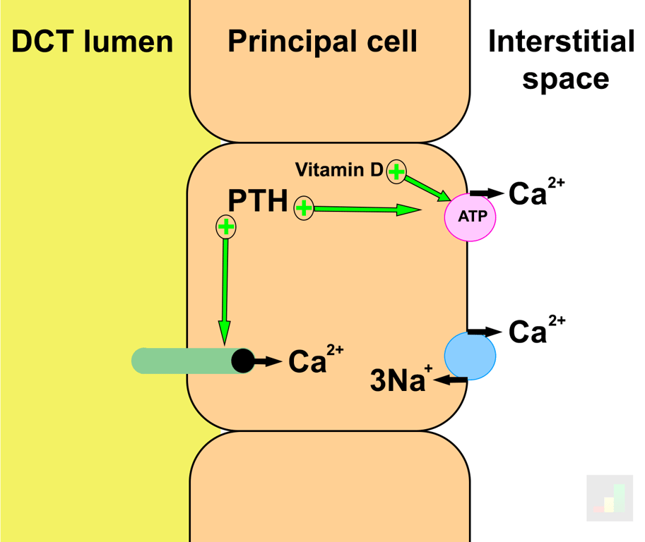 | |
Renal handling of calcium in the distal tubule. Click on image to enlarge