Glomerular filtration
The glomerulus is a capillary network, often described as a tuft or ball of capillaries, supported by mesangium (the space between blood vessels composed of intraglomerular mesangial cells) and surrounded by Bowman's capsule. Blood enters the glomerulus via an afferent arteriole and leaves the glomerulus via an efferent arteriole. The glomerulus is the site of ultrafiltration. The arrangement is shown in the image below:
Mesangial cells are phagocytic and contractile. Contraction may reduce the filtration area.
Bowman's capsule has an inner surface which is covered by specialised epithelial cells called podocytes.
Filtration fraction may be calculated as
An estimated GFR is often used clinically based on serum creatinine, age and sex. An eGFR of 90 mls/min/1.73m2 or above is considered normal.
The relationship is defined by Starlings equation where:
Flow ˆ [Pc - Pi] - σ[Ï€p - Ï€i]
where:
In the glomerulus the approximate figures for hydrostatic pressure are as follows:
In the glomerulus the approximate figures for oncotic pressure are as follows:
Overall there is a net filtration pressure of 10mmHg (35 mmHg - 25 mmHg) driving ultrafiltration
As you can imagine as plasma moves through the glomerulus and is filtered it loses fluid but not protein, the plasma oncotic pressure therefore rises as blood moves through the capillary network form afferent to efferent arteriole. This reduces net filtration pressure near the efferent arteriole. It also means the plasma in the efferent arteriole has a higher oncotic pressure which aids reabsorption when the plasma enters the peritubular capillaries.
Note that the capillary hydrostatic pressure is aided by the unique arrangement of afferent and efferent arterioles. This pressure is reduced by afferent arteriolar constriction and increased by efferent arteriolar constriction. Release of vasoactive agents and adjustments in sympathetic vascular tone allow for differential vasoconstriction and vasodilation of afferent and efferent arterioles. This is important as it allows for autoregulation of renal blood flow and maintenance of a constant GFR over a wide range of blood pressures (90-200 mmHg).
Renal disease, vasoconstrictors* and sympathetic stimulation all reduce GFR.
*Angiotensin II is an exception as it preferentially constricts the efferent arterioles thus works to increase/maintain GFR if blood pressure drops.
Factors affecting filtration barrier permeability
The permeability of the filtration barrier depends mainly on molecular size and molecular charge.
Molecules with a molecular weight below 7000 Da will typically pass freely. These molecules include glucose, amino acids, urea and ions. Permeability decreases as molecular weight increases from 7000 Da to 70,000 Da, molecules weighing more than 70,000 Da typically have insignificant filtration.
Negatively charged particles are typically repelled by the negatively charged proteins that comprise the basement membrane.
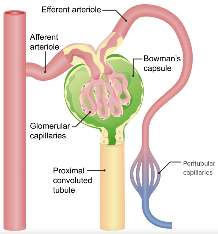 | |
Afferent and efferent arterioles arrangement in the kidney nephron. click on image to enlarge
Mesangial cells are phagocytic and contractile. Contraction may reduce the filtration area.
Bowman's capsule has an inner surface which is covered by specialised epithelial cells called podocytes.
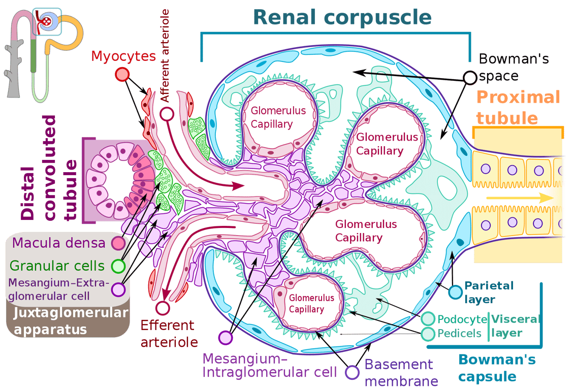 | |
The glomerulus. click on image to enlarge
Ultrafiltration
Ultrafiltration refers to filtration of a liquid, driven by hydrostatic pressure, through a semipermeable membrane that retains colloid particles. Plasma is filtered at the glomerulus in this way.
Ultrafiltration refers to filtration of a liquid, driven by hydrostatic pressure, through a semipermeable membrane that retains colloid particles. Plasma is filtered at the glomerulus in this way.
GFR & filtration fraction
Glomerular filtration rate (GFR) is the most widely used measure of renal function. In healthy human adults GFR is ~ 120 mL/min. Renal plasma flow (RPF) is ~600 ml/min. The proportion of plasma that filters from the glomerulus into the nephron tubule is termed the filtration fraction and this is ~ 20%.Filtration fraction may be calculated as
FF = GFR ÷ RPF
An estimated GFR is often used clinically based on serum creatinine, age and sex. An eGFR of 90 mls/min/1.73m2 or above is considered normal.
Factors determining GFR
GFR depends on the difference between hydrostatic pressure and colloid osmotic pressure in the glomerular capillaries and Bowman's capsule. These forces oppose each other.The relationship is defined by Starlings equation where:
Flow ˆ [Pc - Pi] - σ[Ï€p - Ï€i]
where:
- Pc is the capillary hydrostatic pressure
- Pi is the interstitial fluid (or Bowman's space fluid) hydrostatic pressure
- πp is the plasma protein oncotic (colloidal) pressure
- πi is the interstitial fluid (or Bowman's space fluid) pressure
- σ is Staverman's reflection coefficient
In the glomerulus the approximate figures for hydrostatic pressure are as follows:
- Glomerular capillary hydrostatic pressure = 45 mmHg
- Bowman's capsule hydrostatic pressure = 10 mmHg
- Net hydrostatic force (Pc - Pi) = 35 mmHg
In the glomerulus the approximate figures for oncotic pressure are as follows:
- Plasma oncotic pressure = 25 mmHg
- Bowmans capsule oncotic pressure = 0 mmHg (or close to zero as the ultrafiltrate is virtually protein free)
- Net oncotic pressure = 25 mmHg
Overall there is a net filtration pressure of 10mmHg (35 mmHg - 25 mmHg) driving ultrafiltration
As you can imagine as plasma moves through the glomerulus and is filtered it loses fluid but not protein, the plasma oncotic pressure therefore rises as blood moves through the capillary network form afferent to efferent arteriole. This reduces net filtration pressure near the efferent arteriole. It also means the plasma in the efferent arteriole has a higher oncotic pressure which aids reabsorption when the plasma enters the peritubular capillaries.
Note that the capillary hydrostatic pressure is aided by the unique arrangement of afferent and efferent arterioles. This pressure is reduced by afferent arteriolar constriction and increased by efferent arteriolar constriction. Release of vasoactive agents and adjustments in sympathetic vascular tone allow for differential vasoconstriction and vasodilation of afferent and efferent arterioles. This is important as it allows for autoregulation of renal blood flow and maintenance of a constant GFR over a wide range of blood pressures (90-200 mmHg).
Renal disease, vasoconstrictors* and sympathetic stimulation all reduce GFR.
*Angiotensin II is an exception as it preferentially constricts the efferent arterioles thus works to increase/maintain GFR if blood pressure drops.
Filtration barrier
Fluid from the plasma must through a filtration barrier in the glomerulus to enter Bowman's space and then become tubular fluid that enters the nephron. The filtration barrier has 3 main components:- Endothelial cells of glomerular capillaries
- Glomerular capillary basement membrane
- Epithelial cells of Bowman's Capsule (podocytes)
| Filtration barrier layer | Notes |
|---|---|
| Capillary endothelium | Contains fenestrations (pores) that are 70nm wide that make this layer highly permeable |
| Capillary basement membrane | Consists of 3 layers:
|
| Podocytes | Podocytes contain long extensions called primary processes that engulf capillaries. Pedicels (foot processes) directly contact the basement membrane. Between pedicels are gaps called filtration slits through which filtrate passes. The pedicels ensure the gap between filtration slits is narrow enough to prevent large molecules passing through. Podocytes help maintain the structure of the basement membrane and, like mesangial cells, may be phagocytic and contractile |
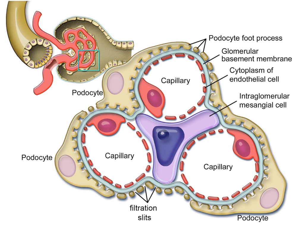 | |
Glomerular filtration barrier. click on image to enlarge
Factors affecting filtration barrier permeability
The permeability of the filtration barrier depends mainly on molecular size and molecular charge.
Molecules with a molecular weight below 7000 Da will typically pass freely. These molecules include glucose, amino acids, urea and ions. Permeability decreases as molecular weight increases from 7000 Da to 70,000 Da, molecules weighing more than 70,000 Da typically have insignificant filtration.
Negatively charged particles are typically repelled by the negatively charged proteins that comprise the basement membrane.
Albumin weighs 69,000 Da and is negatively charged. It is filtered in minute quantities.
As plasma proteins including albumin (the primary plasma protein) are generally large and negatively charged ultrafiltrate is virtually protein free. Ultrafiltrate otherwise has an identical composition to that of plasma.
As plasma proteins including albumin (the primary plasma protein) are generally large and negatively charged ultrafiltrate is virtually protein free. Ultrafiltrate otherwise has an identical composition to that of plasma.