Rectum and anus
On this page
Introduction: rectum and anus
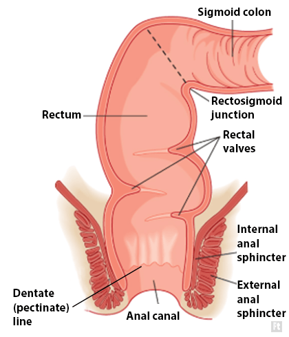
The rectum commences anterior to S3 where the mesocolon ends. It is structurally no different from the sigmoid colon from which it is a continuation.The anal canal is the final part of the gastrointestinal tract. It is approximately 4cm long and is surrounded by internal and external anal sphincters. An anatomical muscular ring (the anorectal ring) marks the junction of the rectum and the anal canal. Histologically there is also a change from the columnar epithelium of the rectum to the stratified squamous epithelium of the anus. The anal canal terminates by piercing the pelvic diaphragm where it becomes the anus.
 | |
Anatomy of the rectum and anus
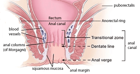 | |
Position of the anal canal
Peritoneal coverings of the rectum
- Upper 1/3 peritoneum to front and sides
- Middle 1/3 peritoneum to front only
- Lower 1/3 devoid of peritoneum
Rectal examination
The following structures are palpable in a normal digital rectal examination:
- Cervix (anterior)
- Prostate (anterior)
- Seminal vesicles (anterior)
- Coccyx (posterior)
Rectum relations
| Posterior relations | Anterior relations (male) | Anterior relations (female) |
|---|---|---|
|
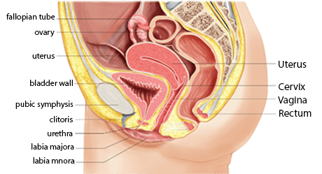 | |
Sagittal section through female pelvis
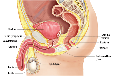 | |
Sagittal section through male pelvis
Innervation of rectum
- Sympathetic from lumbar splanchnic nerves originating from L1-L3. These nerves pass via coeliac, aortic and hypogastric plexuses.
- Parasympathetic from S2-S4 via pelvic splanchnic nerves.
- Pain transmission - visceral afferent (sensory) fibres follow the parasympathetic supply via S2-S4.
Innervation of anal sphincter and perianal skin
- Anal sphincter supplied by S2-S4
- Perianal skin dermatomes innervated in concentric circle pattern by S5, S4, S3
- Anal reflex involves pressing perianal skin with forceps and seeing reflex contraction of anus (external anal sphincter). Involves afferent sensory stimulus via S5 and efferent motor stimulus via S4.
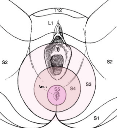 | |
dermatome innervation of the perianal region
note: splanchnic nerves are paired visceral nerves carrying fibres of the autonomic nervous system)
Lymph drainage
- Lower 1/3 rectum / anal canal: Internal iliac nodes and superficial inguinal nodes
- Below the dentate line: superficial inguinal nodes
- Upper and middle 1/3 rectum: Inferior mesenteric nodes via pararectal lymph nodes
- The RCEM syllabus expects candidates to be aware that superficial inguinal nodes may be palpable in disease.
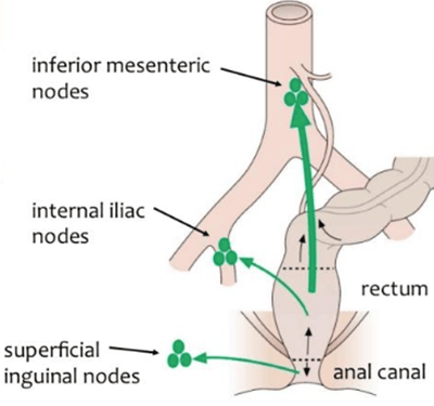 | |
Illustration of lymph drainage of the rectum and anal canal
Arterial supply
Arterial supply is from branches of the inferior mesenteric and internal iliac arteries.
- Superior rectal artery (origin: inferior mesenteric artery)
- Median rectal artery (origin: internal iliac artery)
- Middle rectal artery (origin: internal iliac artery)
- Inferior rectal artery (origin: internal iliac artery)