Eye: Structural anatomy
The eyeball
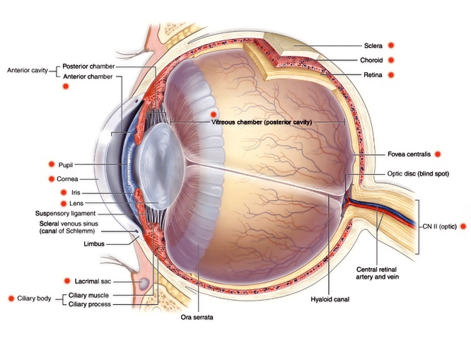
The eyeball has 3 layers or coatings:- Fibrous coat
- Pigmented coat
- Nervous coat
 | |
The fibrous coat: Sclera and cornea
The fibrous coat is the outermost layer and consists of the sclera and cornea. The sclera is a dense fibrous tissue whilst the cornea is transparent and allows light to pass through it.
| Structure | Innervation | Function |
|---|---|---|
| Sclera | Long ciliary nerves (from CN V1) Short ciliary nerves (from CN III) | Maintain shape of eye Protection from trauma |
| Cornea | Long ciliary nerves (from CN V1) Short ciliary nerves (from CN III) | Refraction Transparency Protection |
NOTES: 1. The anterior of the sclera is mostly innervated by the long ciliary nerves whilst the posterior sclera is mainly innervated by the short ciliary nerves.
2. It should be noted that the external part of the eye has an additional outermost layer in the form of the conjunctiva.
The corneal reflex
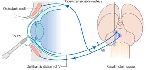
The corneal reflex or blink reflex is required knowledge for the exam. Testing involves:1. Touch the cornea (typically using wisp of cotton)
2. Sensory stimulus transmitted via nasocilliary nerve (branch of CN V1: ophthalmic branch of trigeminal nerve)
3. Impulse reaches trigeminal nucleus in pons
4. Impulse transmitted to facial nerve nucleus in pons
5. Impulses sent via facial nerve to orbicularis oculi to close eyes
 | |
| Wikipedia | |
The pigmented coat
The pigmented coat consists of the highly vascular choroid, the ciliary body and the iris. The ciliary body attaches to the iris and lens and can change their shape via contraction and relaxation of the ciliary muscle. The iris is the pigmented area surrounding the pupil. The iris contains pigmented epithelial cells and muscles that constrict and dilate the pupil (sphincter pupillae and dilator pupillae).
| Muscle | Innervation | Action |
|---|---|---|
| Sphincter pupillae | parasympathetic fibres from CN III (oculomotor) | contstricts pupil |
| Dilator pupillae | sympathetic fibres from CN V1 (long ciliary/nasociliary/ophthalmic branch trigeminal | dilates pupil |
Control of sphincter size is therefor a balance of sympathetic and parasympathetic innervation. The college expect candidates to be aware of this as the basis for the following conditions:
Horner's syndrome
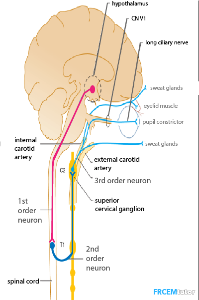
This is the result of loss of sympathetic innervation which arises as the result of damage to the sympathetic nervous system (causative pathology typically either a lesion to brain/neck or lung apex). The result is seen clinically as a unilateral small pupil, ptosis and anhidrosis.The pathway that provides the sympathetic innervation to the eye consists of three neurons referred to as 1st, 2nd and 3rd order neurons. Horner's syndrome can be the result of disturbance to any of these three neurons and can be classified as 1st, 2nd or 3rd order Horner's syndrome as a result. 1st order neurons have their cell bodies in the hypothalamus and their axons run through the brainstem and cervical cord. These 1st order neutrons synapse with second order neurons in the thoracic spinal cord. The axons of the second order neurons leave the spinal cord and join the thoracic sympathetic trunk then ascend to the superior cervical ganglion. Here the second order neurons synapse with the 3rd order neurons. The 3rd order neurons carry impulses from the superior cervical ganglion to the effector muscles.
 | |
Argyll-Robertson pupil
Bilateral small pupils that don't react to light but do constrict during accommodation. Pathophysiology is believed to be the result of bilateral damage to the pretectal nuclei in the midbrain. The sign is strongly associated with neurosyphilis.
The nervous coat: Retina
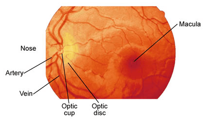
The retina is the light sensitive layer and contains the photoreceptor cells (rods and cones).The optic nerve enters the retina, accompanied by the central artery, at the optic disc which corresponds clinically to the blind spot.
The macula lies temporal to the optic disc. It is seen on fundoscopy as a darker/yellow spot. The macula contains a high density of light receptors. The depressed central area of the macula is termed the fovea (or fovea centralis) and contains the highest density of cone cells. As a result this area has the greatest visual acuity. The macula and fovea receive their blood supply from the choroid rather than directly from the central retinal artery.
 | |
Chambers of the eye
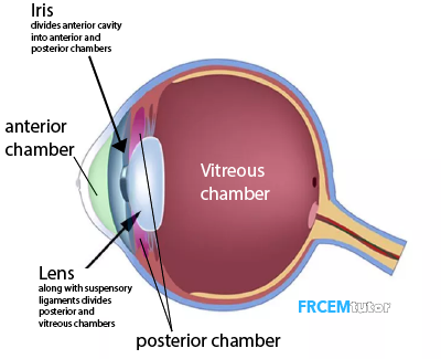
The naming of the chambers/cavities or segments of the eye can be confusing and isn't helped by several texts and online resources getting this incorrect. It isn't helped either that the posterior chamber isn't the most posterior of the chambers!
To clarify there are three chambers.
| Chamber | Position | Fluid |
|---|---|---|
| Anterior chamber | Immediately behind/posterior to cornea Anterior to iris | aqueous humour |
| Posterior chamber | Posterior to the peripheral part of the iris Anterior to lens, suspensory ligament of the lens and the ciliary processes | aqueous humour |
| Vitreous chamber | Posterior to lens | vitreous humour |
 | |