Paranasal Sinuses
Overview
The paranasal sinuses are paired structures and include the following:
- Maxillary sinuses
- Ethmoidal sinuses
- Frontal sinuses
- Sphenoidal sinuses
Key Points
- They are lined with ciliated pseudostratified columnar epithelium (respiratory epithelium)
- The maxillary sinuses are the largest of the paranasal sinus group
- The paranasal sinuses are innervated by branches of the trigeminal nerve
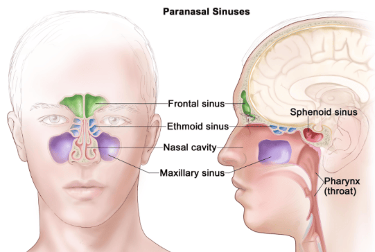 | |
| Wikipedia | |
Mucus Drainage
- The frontal and maxillary sinuses drain into the middle meatus
- The sphenoid sinuses drain into the sphenoethmoidal recess
- The ethmoidal sinuses drain into the middle and superior meatus
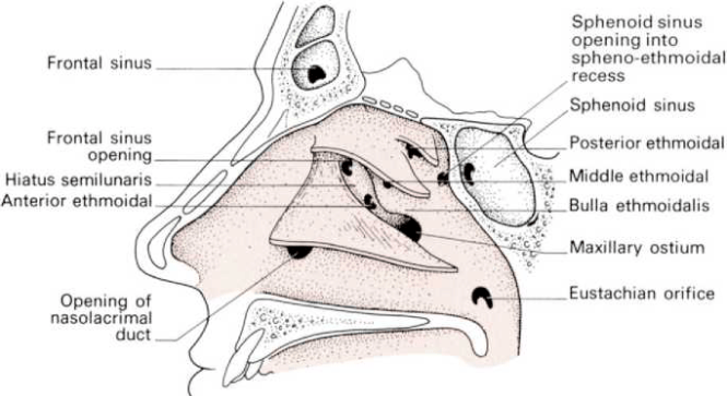 | |
Development & Expected Knowledge
- The college expect candidates to be able to identify the sinuses on plain XRAY and also have an understanding of their development from age 7 onwards
- Enlargement is via excavation of bone by pneumatic diverticula
| Sinus | First Detected | Development |
|---|---|---|
| Ethmoid | Present at birth | grow and pneumatize until the age of 12 |
| Maxillary | Present at birth | 2nd growth phase between 6-12 inferiorly towards teeth |
| Sphenoid | Detected at 3-4 years | From age 7 extends posteriorly, completing growth in teenage years |
| Frontal | Detected at 7-12 years | Growth completed by age 20 |