Myocarditis
On this page
Definition
Myocarditis refers to inflammation of the myocardium. It may be acute or chronic, and can mimic myocardial infarction with chest pain and ECG changes, but is distinguished by non-ischemic injury.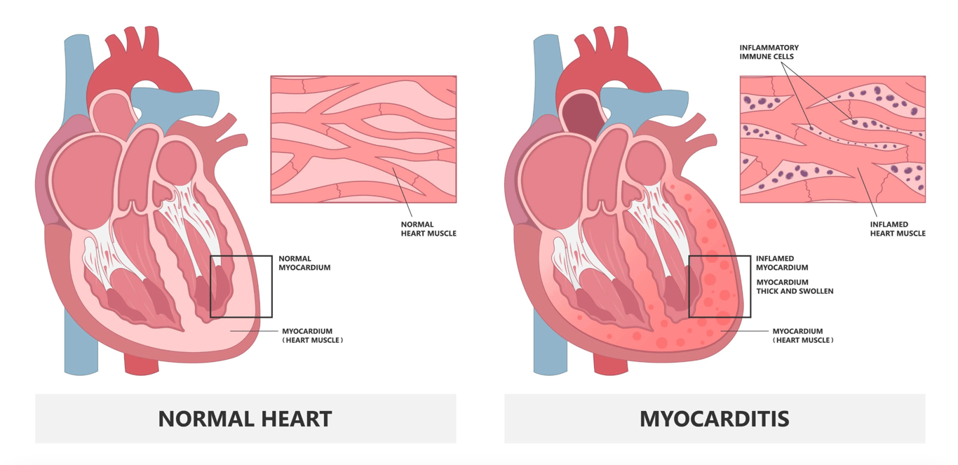 | |
Click on image to enlarge
Epidemiology
True incidence is difficult to gauge as the condition is often underdiagnosed. In England, myocardial inflammation accounted for ~0.04% of hospital admissions.
More common in Men with male to female ratio of 1.5:1
Aetiology
Infectious causes (most common):- Viruses: Coxsackievirus, adenovirus, parvovirus B19, enteroviruses, Civid-19, HIV, EBV, hepatitis viruses
- Bacteria: e.g., diphtheria
- Parasites, fungi, spirochetes (in endemic or immunocompromised patients)
- Autoimmune conditions (SLE, sarcoidosis)
- Hypersensitivity / eosinophilic myocarditis
- Giant cell myocarditis
- Drugs: anthracyclines, cocaine, cytotoxic agents
- Radiation, heavy metals, chemicals, electric shock, hyperthermia
- Coxsackie virus is the most common viral cause of myocarditis in the UK, Europe and the USA
- Worldwide Diptheria is the most common bacterial cause
- Lithium, cocaine and several chemotherapy agents may cause myocarditis via a direct cytotoxic effect.
- Several drugs may also cause myocarditis via hypersensitivity reactions.
Clinical features
There is a wide spectrum of presentations from asymptomatic to fulminant myocarditis with rapid cardiac failure.
Symptoms include:
- Fatigue
- Chest pain (~35%)
- Dyspnoea
- Palpitations (arrhythmias)
- Fever
- Heart failure signs (oedema, paroxysmal nocturnal dyspnoea etc)
- Soft S1, S4 gallop
Investigations
| Investigation | Findings |
|---|---|
| ECG | Non-specific abnormalities: ST elevation/depression, T wave inversion, AV block, supraventricular arrhythmias |
| Blood tests | Elevated troponin, CK; inflammatory markers (CRP, ESR); full blood count, renal & liver panels |
| CXR | Cardiomegaly, pulmonary congestion (if heart failure) |
| Cardiac MRI | Tissue inflammation, edema, scar; differentiates myocarditis from infarction |
| Endomyocardial biopsy | Gold standard; definitive histology; used in selected cases |
Differential diagnoses
- Acute coronary syndrome (MI)
- Congestive heart failure from other causes
- Pulmonary edema / pneumonia
- Other chest pain causes (PE, pericarditis)
Management
- 1. Supportive therapy: inotropic support, heart failure management
- 2. Severe cases: ICU admission, mechanical circulatory support if needed
- 3. Treat underlying cause: antivirals, antimicrobials if applicable
- 4. Immunosuppression / steroids / IVIG: only in specific cases (giant cell, eosinophilic myocarditis)
- 5. Anticoagulation: if thromboembolism risk is high (AF, ventricular thrombus)
- 6. Restrict physical activity post-acute phase for myocardial recovery
Complications
- Dilated cardiomyopathy and chronic heart failure
- Cardiogenic shock
- Arrhythmias and sudden cardiac death
- Recurrent myocarditis
Prognosis
Prognosis is variable ranging from complete recovery to chronic heart failure or death
Prognostic factors: left ventricular dysfunction, hemodynamic compromise, arrhythmias, myocardial inflammation/scarring
Summary
| Feature | Key Points |
|---|---|
| Definition | Inflammation of myocardium; may mimic MI |
| Common Causes | Viral infections, autoimmune, drugs/toxins |
| Presentation | Fatigue, chest pain, dyspnoea, palpitations, fever |
| Investigations | ECG, troponin, CXR, cardiac MRI, biopsy |
| Treatment | Supportive, treat underlying cause, ICU if severe |
| Complications | Heart failure, cardiogenic shock, arrhythmias, recurrent myocarditis |
| Prognosis | Highly variable; depends on LV function and severity |