Lung anatomy
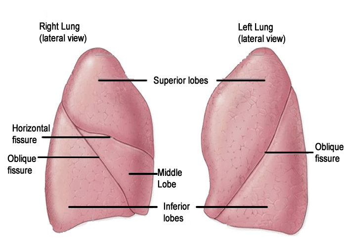
The lungs are divided into lobes by the fissures. The right lung is divided into 3 lobes by horizontal and oblique fissures. The left lung is divided into two lobes by an oblique fissure as pictured below:
Image illustrating lung lobes
Surface anatomy
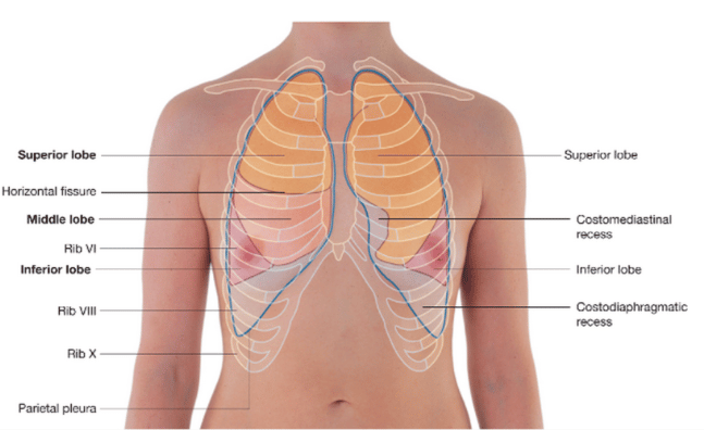
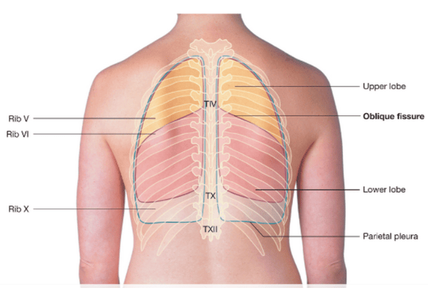
You are expected to be familiar with the surface anatomy of the lungs, fissures and hila. The images below demonstrates the key surface anatomy
Image illustrating lung surface anatomy
Lung surface anatomy:
Fissures:
Hila:
The lung hila correspond to the level of the 3rd and 4th costal cartilages anteriorly and T5-T7 vertebral level posteriorly. Relative to the midline they are approximately 2.5cm lateral to the sternal edge anteriorly or 5cm lateral to the thoracic spines/midline posteriorly.
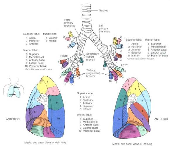
Image illustrating bronchopulmonary segments
Image illustrating lung lobes
 | |
| Wikipedia | |
Surface anatomy
You are expected to be familiar with the surface anatomy of the lungs, fissures and hila. The images below demonstrates the key surface anatomy
Image illustrating lung surface anatomy
 | |
| Wikipedia | |
 | |
| Wikipedia | |
Lung surface anatomy:
- Apex: found posterior to medial 1/3 clavicle and extends 2.5cm above the clavicle.
- Medial border: posteriorly extends from C7 spinous process to T10 vertebra 4cm lateral from midline
- Inferior border: midclavicular line - 6th rib, midaxillary line - 8th rib and 10th rib adjacent to vertebral column
- Inferior border: posteriorly T10 vertebral level
Fissures:
- Horizontal fissure (right lung only): extends laterally in horizontal line from 4th costal cartilage to meet oblique fissure in midaxillary line
- Oblique fissure: The oblique fissure runs from the spinous process of T3 to the 6th costal cartilage anteriorly
Hila:
The lung hila correspond to the level of the 3rd and 4th costal cartilages anteriorly and T5-T7 vertebral level posteriorly. Relative to the midline they are approximately 2.5cm lateral to the sternal edge anteriorly or 5cm lateral to the thoracic spines/midline posteriorly.
Image illustrating bronchopulmonary segments
 | |
| Wikipedia | |