Liver surface anatomy
Surface anatomy
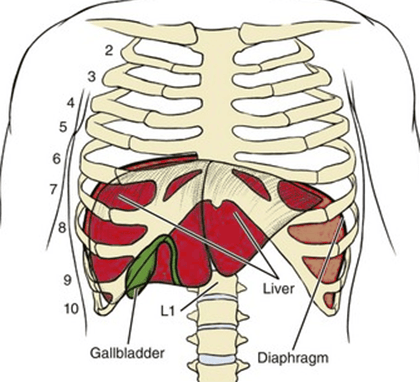 | |
Diagram illustrating approximate position of liver. Note the liver moves with respiration
Superior surface
- Reaches the 5th rib on the right
- Reaches 6th rib 7cm from midline on the left
- Just below the 10th rib on the right side
- Inferior border can be marked by line from 10th costal cartilage right midaxillary line to 6th rib left midclavicular line.
- Laterally the superior border of the liver reaches the 6th rib in the mid-axillary line
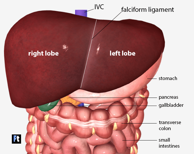 | |
Anterior view of the liver
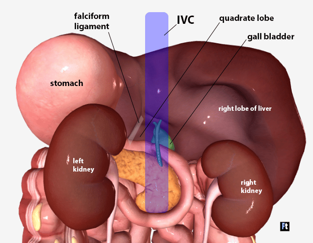 | |
Posterior view of the liver. Note the stomach obscures the left lobe of the liver in this image. The stomach has been removed on the illustrations below
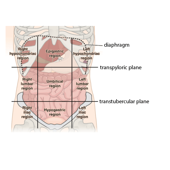 | |
The 9 region abdominal anatomical scheme uses the vertical midclavicular lines and horizontal transpyloric and transtubercular planes to divide the abdomen into 9 regions
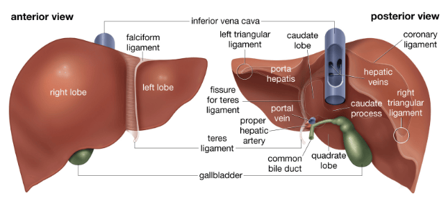 | |
Anterior and posterior views of the liver
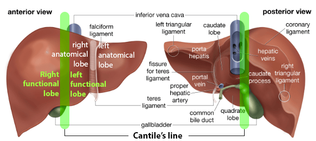 | |
The green lines highlight Cantlie's line, a vertical line drawn between the IVC superiorly and gallbladder fossa. Cantile's line separates the functional lobes where as the falciform ligament separates the anatomical lobes