Anaemia: classification and overview
Definition
Anaemia (anemia) is normally defined by a haemoglobin (Hb) value below the reference range for age and sex, commonly defined as more than 2 standard deviations below the mean.The RCEM quote the normal Hb reference range for adults as 11.5 - 16.6g/dl and normal MCV range as 80-96 fl, though most UK hospitals use an MCV range of 80-100 fL.
Classification
Anaemia is most commonly classified based on the red blood cell volume (MCV) into:- Microcytic (MCV < 80 fL)
- Normocytic (MCV 80-100 fL)
- Macrocytic (MCV > 100 fL)
Mean cell haemoglobin (MCH) content may also be used to classify anaemia as hypo, hyper or normochromic.
Macrocytic anaemias are broadly further classified into two main types: megaloblastic and non-megaloblastic.
Megaloblastic anaemia is often caused by folate or vitamin B12 deficiencies. These deficiencies impair DNA synthesis and lead to cell abnormalities characterised by the presence of large, immature, and dysfunctional red blood cell precursors (megaloblasts) in the bone marrow and macrocytic (large) red blood cells in the peripheral blood.
Megaloblastic anaemia is often caused by folate or vitamin B12 deficiencies. These deficiencies impair DNA synthesis and lead to cell abnormalities characterised by the presence of large, immature, and dysfunctional red blood cell precursors (megaloblasts) in the bone marrow and macrocytic (large) red blood cells in the peripheral blood.
Causes
Red blood cells are produced in the bone marrow in response to the hormone erythropoietin (EPO). In adults, EPO is primarily produced by peritubular interstitial cells in the kidney in response to hypoxia (low oxygen levels). The kidneys are responsible for approximately 90% of EPO production in adults, with the liver contributing the remaining 10%. In the fetus, however, the liver is the main site of EPO production.Anaemia may result from various causes, including deficiencies in nutrients essential for haemoglobin or red blood cell synthesis (such as iron, vitamin B12, or folate), inadequate or impaired EPO production, or increased red cell loss or destruction (as seen in haemolysis or bleeding)
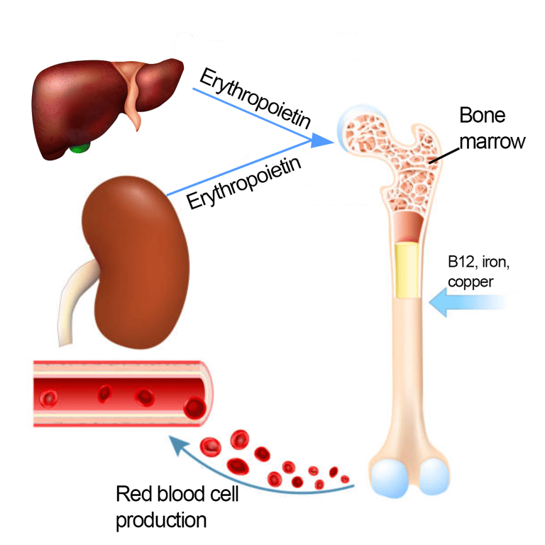 | |
Red cell production. Click on image to enlarge
Causes of anaemia
| Anaemia Type | MCV Range (fL) | Common Causes |
|---|---|---|
| Microcytic | < 80 |
|
| Normocytic | 80–100 |
|
| Macrocytic | > 100 |
|
*May result in normocytic or microcytic anaemia
Physiological response to anaemia
When anaemia develops, the body initiates several compensatory mechanisms to maintain adequate oxygen delivery to tissues. The kidneys detect reduced oxygen-carrying capacity in the blood and respond by increasing production of erythropoietin (EPO), a hormone that stimulates red blood cell production in the bone marrow. The cardiovascular system also adapts: heart rate and cardiac output increase, helping to circulate the available oxygen more efficiently. Additionally, there is a rightward shift in the oxygen–haemoglobin dissociation curve (via increased 2,3-BPG levels), which facilitates oxygen release from haemoglobin to tissues. Over time, increased respiratory rate and plasma volume expansion also help to improve oxygen delivery. These responses can temporarily compensate for mild to moderate anaemia, but they become insufficient in severe or chronic cases, where symptoms like fatigue, breathlessness, and tachycardia become more prominent.Investigations
Investigations are typically guided by the type of anaemia and the clinical context. For example, iron deficiency anaemia in postmenopausal women or men raises suspicion for gastrointestinal blood loss, and is often investigated with endoscopy to rule out malignancy or occult bleeding. In contrast, megaloblastic anaemia typically prompts assessment of vitamin B12 and folate levels and may require further haematological evaluation if a primary bone marrow disorder is suspected.Investigations may include:
- Full blood count (FBC, also known as complete blood count or CBC)
- Iron studies: ferritin, transferrin and TIBC
- B12, Folate
- Peripheral blood film
- Liver function test
- Urea & electrolytes
- Thyroid function tests
- Endoscopy
- Haematological investigations e.g. bone marrow biopsy, flow cytometry, genetic testing, heamoglobin electrophoresis.
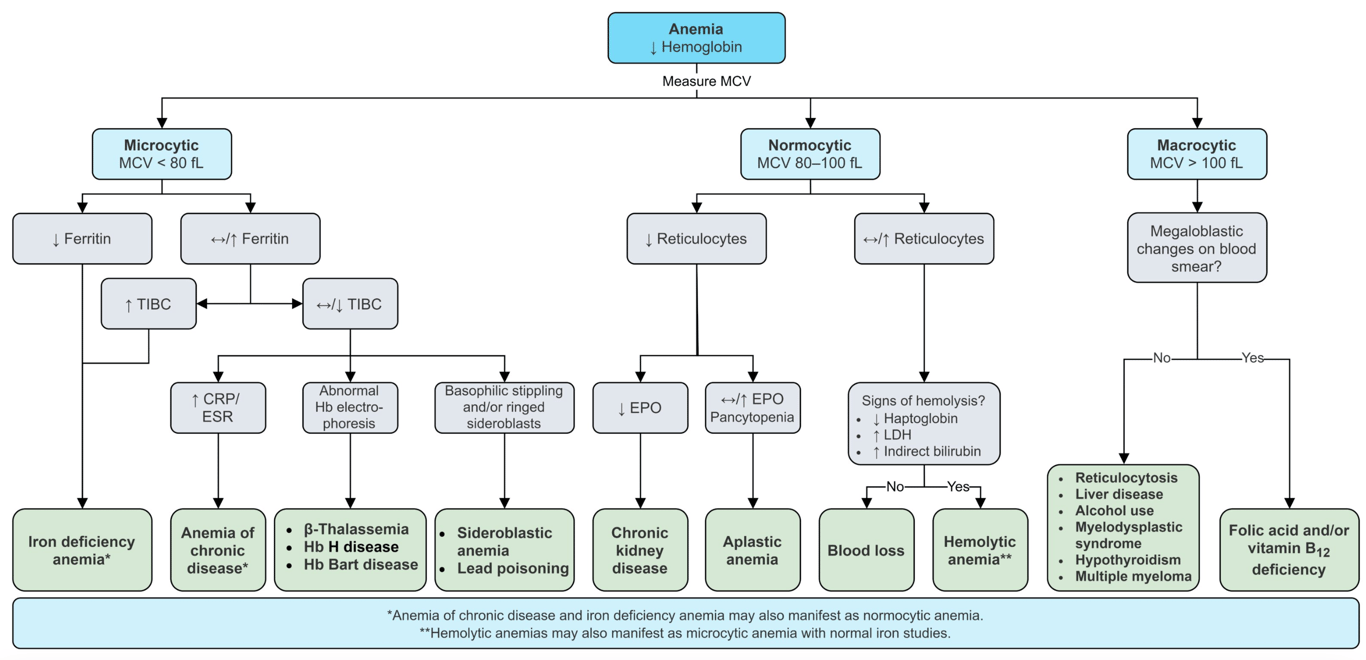 | |
Classification and investigation of anaemia. Click on image to enlarge
Signs and symptoms
- Fatigue
- Pallor
- Shortness of breath
- Reduced exercise tolerance
- May precipitate angina in those with pre-existing coronary artery disease
- Tachycardia
- Palpitations
- Headache
- Cognitive dysfunction
- Restless leg syndrome
- Dizziness or lightheadedness
- Weakness
- Dysgeusia (altered sense of taste)
- Irritability
- Pruritus
- Sore tongue (glossitis)
- Tinnitus
- Angular stomatitis
- Nail changes (koilonychia in iron deficiency, pale nails)
- Jaundice may be present in heamolytic anaemia
- Peripheral neuropathy or gait disturbance in B12 deficiency
Management
Once the underlying cause has been identified management is to correct underlying deficiencies with supplementation e.g. iron , B12 or folate supplementation and treat the underlying cause.
In combined B12 and folate deficiency, B12 should be replaced first to prevent worsening neurological damage.