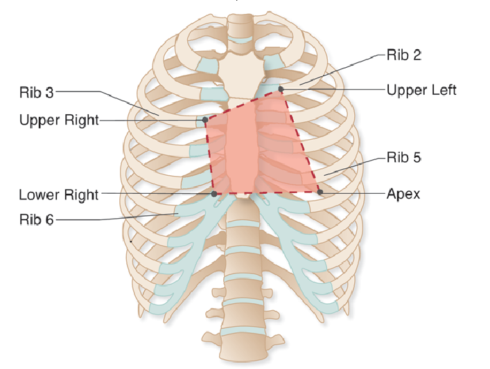
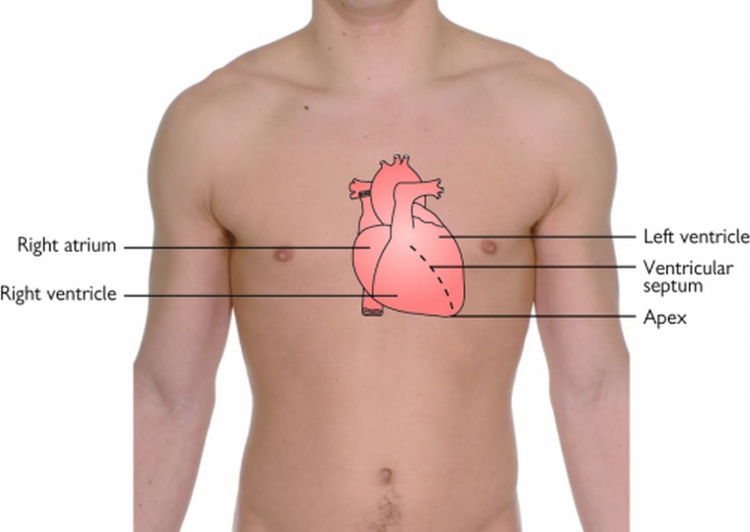
The diagrams below show the surface markings of the heart and the orientation of the heart chambers
Image illustrating borders of the heartThe college expects you to appreciate the cardiac structures making up the borders and surfaces of the normally-orientated heart. It is worth remembering the 4 corners of the heart (see image above and table below)
| Border | Structure | Landmarks |
|---|
| Right border | Right atrium | - Upper: 3rd right costal cartilage
- Lower: 6th right costal cartilage
|
| Left border | Left ventricle & left atrial appendage | - Upper: 2nd left costal cartilage
- Lower: 5th left ICS midclavicular line (apex)
|
| Superior border | Both atria & roots of great vessels | - Right: 3rd right costal cartilage
- Left: 2nd left costal cartilage
|
| Inferior border | Right ventricle & apex of left ventricle | - Right: 6th right costal cartilage
- Lower: 5th left ICS midclavicular line (apex)
|
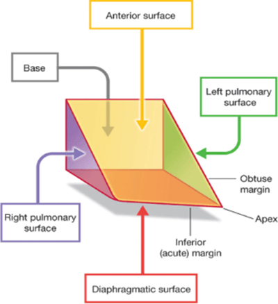
In 3D terms the heart is sometimes described as being a pyramid structure with the apex pointing down and to the left.
Image illustrating heart surfaces| Surface | Structure |
|---|
| Sternocostal (anterior) | Right ventricle (2/3) and right atrium (1/3) |
| Diaphragmatic (inferior surface) | Right and left ventricle |
| Posterior (base of pyramid) | Left atrium |
| Pulmonary (left) | Left ventricle |
| Pulmonary (right) | Right atrium |
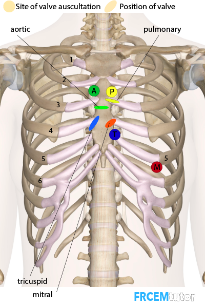
| Valve | Auscultation site | Surface marking / anatomical position |
|---|
| Aortic | 2nd ICS right sternal edge | 3rd ICS retrosternally |
| Pulmonary | 2nd ICS left sternal edge | 3rd rib left sternal edge |
| Tricuspid | 4th ICS left sternal edge | 4th ICS right sternal edge |
| Mitral | 5th ICS left midclavicular line | 4th rib left sternal edge |