Gout
On this page
Background
Gout is an inflammatory arthritis caused by deposition of monosodium urate crystals in joints and peri-articular tissues due to persistent hyperuricaemia. It typically presents with sudden, severe monoarticular pain and swelling. Acute flares often reach maximal intensity within 12 hours and are interspersed with asymptomatic periods. The first metatarsophalangeal joint (1st MTP) is involved in approximately 50% of first attacks. Other commonly affected joints include the ankle, knee, wrist, and fingers.Epidemiology
Gout is the most common inflammatory arthritis. Prevalence has risen in recent decades and is higher in men and with advancing age; UK prevalence estimates are approximately 1–3%. Risk increases with co-morbidities such as obesity, chronic kidney disease, hypertension, and metabolic syndrome.Causes & risk factors
- Persistent hyperuricaemia: overproduction or under-excretion of uric acid.
- Diet and lifestyle: high alcohol intake (especially beer), high purine foods, sugary drinks, obesity.
- Medications: thiazide or loop diuretics, low-dose aspirin, ciclosporin, pyrazinamide.
- Comorbidities: chronic kidney disease, hypertension, heart failure, metabolic syndrome.
- Genetic predisposition and ageing.
Clinical presentation
- Pain: typically sudden, severe, often nocturnal, reaching maximal intensity within 12 hours.
- Swelling, erythema, and tenderness at the affected joint.
- Pattern: typically monoarticular in early disease, commonly 1st MTP (~50%), but may affect ankle, knee, wrist, or fingers.
- Recurrent flares can lead to chronic joint damage and tophi if untreated.
- Systemic features are usually absent, but fever and malaise may occur in severe attacks.
Diagnosis & assessment (ED perspective)
- Synovial fluid aspiration is the gold standard diagnostic test: polarised light microscopy showing needle-shaped, negatively birefringent crystals (blue when parallel to slow axis). However this is NOT usually performed in the ED.
- Serum urate levels help with long-term management but may be normal during an acute flare.
- Blood tests: inflammatory markers such as CRP are typically elevated
- X-ray: may show early joint effusion, in chronic gout 'punched-out' erosions with sclerotic margins, overhanging edges, eccentric erosions and/or soft-tissue tophi may be seen. Joint space is typically preserved until late disease. Note no periarticular osteopenia (contrast with RA).
- Ultrasound or dual-energy CT can detect urate deposits if aspiration is not feasible.
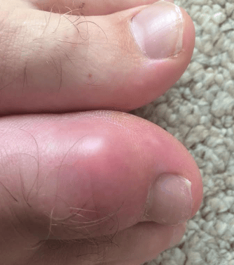 | |
Photograph of a patient with gout affecting the hallux. Click on image to enlarge
Acute Management
- NSAIDs: first-line if no contraindication; co-prescribe PPI if required.
- Colchicine: alternative if NSAIDs contraindicated. Adjust dose for renal function.
- Oral corticosteroids: are another alternative e.g., prednisolone 30–35 mg once daily for 3–5 days, or intra-articular steroids for single-joint flares (though this is rarely done in the ED)
- Supportive care: analgesia, elevation, joint rest.
- Rule out septic arthritis: aspirate joint if infection cannot be excluded.
Preventing gout
- Urate-lowering therapy (ULT) is indicated for recurrent flares, tophi, chronic gouty arthritis, uric acid nephrolithiasis, or CKD stage 3+. Allopurinol is first choice (usually initiated by primary care rather than ED). Alternative ULT includes febuxostat and uricosurics which may be used if allopurinol-intolerant or refractory.
- Flare prophylaxis: low-dose colchicine or NSAID when starting or increasing ULT. Continue ULT during acute attacks.
- Lifestyle modification: low purine diet, weight loss, alcohol reduction and reduce sugary drinks.
- Review medications and consider stopping medications that exacerbate gout.
Medications that increase risk of gout:
- Diuretics: thiazides (e.g., bendroflumethiazide, hydrochlorothiazide) and loop diuretics (e.g., furosemide) reduce renal uric acid excretion.
- Low-dose aspirin: can reduce uric acid excretion.
- Immunosuppressants: ciclosporin.
- Anti-TB agents: pyrazinamide, sometimes ethambutol.
- Other drugs: nicotinic acid (high-dose niacin), cytotoxic drugs (less common).
Complications & Prognosis
- Chronic untreated hyperuricaemia can cause tophaceous gout, joint destruction, chronic pain, and deformity.
- Renal complications: uric acid nephrolithiasis and urate nephropathy.
- With effective long-term ULT and lifestyle modification, prognosis is good, flares are reduced, and tophi may resolve.